material upon the tooth, where normally
smooth, non-irritating cuticle exists, soon causes inflammation;
at first only microscopic in extent. Minute and microscopic
quantities of inflammatory exudate and pus cells are poured out
through the inflamed tissue into the gingival crevice, and these
tend to promote the growth of microorganisms and increase of the
concretion. As the foreign material on the tooth increases and
advances further into the gingival crevice, the soft tissues
attached to the tooth are forced back by the accompanying
inflammation and ulceration. Gradually and almost imperceptibly
a larger and larger portion of the tooth is exposed as the
gingival margin moves further away from the occlusal level. The
gum "recedes."
Many factors influence the progress of the
lesions at different locations about a given tooth, about
different teeth in the same individual and in different
individuals. For our present purpose of prevention of the
disease (lesions) and prevention of further progress of lesions
that already exist, it is only necessary to adopt and direct
effective measures against the conditions at and within the
gingival crevice. The disease process involving the more remote
tissues—periodontal membrane, alveolar bone—rapidly subsides as
soon as the local lesions consisting of inflamed, suppurating
and broken surface of the epithelial tissue of the gum within
the crevice, subside and disappear.
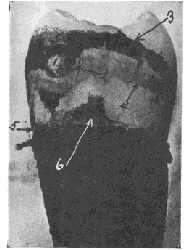 |
Fig. 10.
Proximal of extracted tooth stained with crystal
violet showing biconcave disc of bacterial material
surrounding contact area, and heavy film which
extended from the gingival border (gingival crevice)
occlusalward. Contact point (1). Disc of bacterial
film material surrounding contact point (2). Heavy
bacterial material on area protected from functional
friction (3). Epithelial cells remaining attached to
tooth (4). Location of cemento-enamel junction (5).
Some epithelial attachment tissue retained on tooth
(6). |
From the earliest stage and continuously as
the lesion progresses, the tooth surrounded by this concretion
and bacterial material adhering to it within the gingival
crevice is, in effect, a foreign body infected with many
different kinds of bacteria. It is a suppurating lesion
constantly exposed to invasion by any and all of the many
different kinds of bacteria within the mouth which are capable
of growing in such an environment.