Personal Oral Hygiene
Helping people clean and keep their teeth since 1961!
Personal Oral Hygiene
Helping people clean and keep their teeth since 1961!
|
|
In order to devise and adapt personal oral hygiene measures for prevention of the formation of the material upon the tooth which causes and promotes progress of the disease, correct conception of the nature of this material is necessary. We have already seen that the bacterial film (tartar) on the surface of a tooth above the gingival margin at the more protected places where it can accumulate and where it is not dislodged by functional friction (Figures 10,11), consists largely of a thick pad or pile of long rod and filamentous bacteria, one end of which is attached to the enamel cuticle. The other end extends outward to the surface of the film pack where there are usually an abundance of other bacteria of many different kinds.
The gingival margin must rest against this bacterial mass of foreign material which causes irritation, inflammation and suppuration. As the foreign material on the tooth builds up and advances into the gingival crevice (Figure 12) the inflammatory exudate there offers favorable environmental conditions and nutritive material for the growth of other types of microorganisms that do not grow outside of the crevice. They are organisms, such as certain leptotrichia, actinomyces, spirochetes. ameba, etc., which prefer or require the more or less anaerobic conditions, inflammatory tissue exudate, blood and pus present in such diseased gingival crevices. After a lesion, although small, is well established at any particular location and any time thereafter during the advancement of the lesion, the surface of the tooth within the crevice against which the inflamed gingival surface rests, has more or less hard calculus on it at most areas. The inner border of such calculus approaches but usually does not quite reach the zone of disintegrating epithelial attachment cuticle,(1) a landmark that can be seen on extracted teeth and indicates the exact location of the outer border of the epithelial attachment. Superimposed upon and attached to the calculus and any part of the tooth on which there is none, within the gingival crevice, there is a pad or pile of soft bacterial material. This bacterial film consists largely of closely packed parallel long rod and filamentous forms, one end of which is attached to the calculus or the tooth from which the rod or filament extends outward toward the surface of the pad against which the inner inflamed surface of the gum rests. At the surface of the bacterial film within the crevice there are growing ends and fruiting heads of the rods and filaments composing the pad, and among these more or less other bacteria that invade the lesion from the mouth. Among elements making up the film pad on the tooth within the gingival crevice perhaps the stems and fruiting heads of Leptothrix falciformis are the most noticeable and the most constant. This organism was first described in material from around teeth by Beust(17) in 1906 and 1908. I (5) have called attention to the fact that the habitat of Endameba buccalis is among the stems, branches and fruiting heads composing this film pack. |
 Fig.
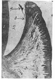
11. (above) Proximal of
extracted molar stained to show bacterial film.
Contact area (1). Heavy bacterial film (2).
Epithelial cells remaining attached to tooth (3).
Location of cemento-enamel
junction (4).
Fig.
11. (above) Proximal of
extracted molar stained to show bacterial film.
Contact area (1). Heavy bacterial film (2).
Epithelial cells remaining attached to tooth (3).
Location of cemento-enamel
junction (4).