The
Necessary Personal Oral Hygiene
For Prevention of Caries and Periodontoclasia*
|
|
by Charles C. Bass,
M.D |
Page 2-continued
Sometimes a small broken down area (cavity) may
be observed in a larger area of partially decalcified enamel
which still holds its form. It will be observed that most small
to medium size cavities have more or less unbroken chalky enamel
about them. (Figures 2, 3).
|
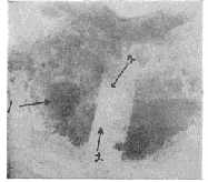
Fig. 2 Area on proximal of molar with cavity
(1) and chalky enamel (2). Two parallel cuts were made
through the cuticle to include the edge of the chalky
enamel. Specimen in acid I minute, rinsed and then
stained with crystal violet. Strip of loose cuticle
removed, exposing some of "white spot" area. Clear strip
from which cuticle was removed contrasts well with
stained cuticle and bacterial film still in place on
either side.
|
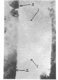
Fig. 3. Higher magnification of area on Fig.
10, showing chalky enamel extending outward from beneath
cuticle on left side which was retained in place.
|
No cavity ever forms except as a result of
breaking down of this earlier stage decalcified enamel. The
early stage, partial decalcification, therefore always precedes
cavity formation. Prevention of cavity formation and its
consequences can be accomplished only by recognition of the
etiological conditions at the location where the earlier partial
decalcification occurs and by application there of effective
measures for preventing or minimizing those conditions.
The Enamel Cuticle In
Relation To The Early Stages of Caries
It has been shown (2)
that the enamel cuticle bears an important-relationship to the
early stage of caries. The enamel cuticle is an extremely thin
keratin-like, transparent membrane covering the entire enamel
surface at all times. It is thinner over areas where it is
repeatedly worn by functional or other friction than in other
areas where it is not exposed to such friction. However it is
extremely thin in such areas also. In view of some confusion and
conflicting opinion as to the continued presence throughout life
of an enamel cuticle, it may be worthwhile to give here a simple
procedure whereby anyone who is interested can clarify the
matter for himself. Again this can be done without the aid of
microscopic laboratory equipment or experience.Place a tooth
specimen in the 10 per cent HC1 for
one minute; remove gently and dip in water for a moment to
reduce the acid; place in 0.5 per cent crystal violet solution
(crystal violet 0.5 gm in water 100 cc) for one minute or less;
again dip in water to remove excess of stain. Now observe the
loosened cuticle with the tooth immersed in a shallow dish of
water in which the membrane may be teased off with some suitable
delicate instrument (No. 2 or No. 7 Clevdent or S. S. White
Explorer) and manipulated in the water. The cuticle itself is
slightly stained and the bacterial film upon it is heavily
stained. When floating in the water the membranous nature of
this material from the surface of the enamel is readily
recognized. One who examines a few specimens in this simple way
knows, of his own knowledge, that an enamel cuticle is
continuously present on teeth. This enamel cuticle is of
interest in relation to caries because the bacterial film over
the early stage lesion is firmly attached to the cuticle (Figure
4) and because the acid or acids which cause the first partial
decalcification there must pass through the intact membrane to
reach the enamel.
Continued...
|